

Kerr at 150: A Community Mosaic
Celebrating 150 years of the magneto-optical Kerr effect
Announcement:

All submissions to be displayed at the upcoming MMM 2026 @ Honolulu, Hawaii!
Thanks to the generous support of evico magnetics every submission to Kerr at 150: A Community Mosaic received before the deadline will be printed and displayed at the upcoming MMM 2026. This means your contribution to the mosaic won't just live online — it'll be part of a physical exhibit for the community to see in person.
About
2026 marks 150 years since John Kerr's discovery of the magneto-optical Kerr effect (MOKE) that revolutionized how we image and understand magnetism. To mark this milestone, we invite the magnetics community to contribute your Kerr images — from domain walls to skyrmions, thin films to bulk crystals, and loops that tells of magnetic responses — and together build a snapshot of where the effect stands today.
_edited_edited_edit.jpg)
Crafting the Mosaic
We invite any researcher in the magnetics community to submit your Kerr images — beautiful, surprising, scientifically significant, or simply ones that meant something to your work.
Along with your image(s), tell us your story. What were you measuring, and why? What were the conditions? Did a particular MOKE measurement change the direction of your research, open a new question, or finally confirm something you had long suspected? There are no strict rules — captions, context, and backstory are all welcome. You are encouraged to submit more than one image.
We welcome all submissions related to the Kerr effect (not restricted to MOKE imaging). Feel free to submit your MOKE imaged domains, any measurements related to the effect, your MOKE-measured loops, your set-ups with your research groups, and more! Anything related to the effect to tell of how it has impacted the community.
Submissions close on 15 June 2026 (extended & closed). Submissions received beyond 15 June 2026 will not be considered for the event.
Please note: by submitting, contributors confirm they hold the right to share the image. SiM takes no responsibility for the accuracy, completeness, or publication status of submitted content. Submitting unpublished data remains the sole responsibility of the contributor.
The Gallery
Explore the compilation of all submissions and their stories. Make use of the left/right navigation arrows to see the multiple images that involve the Kerr effect.
Cast your vote for your favorite entries here:
CDF-01


Christoph Durner
Fraunhofer IPMS CNT
PhD Candidate
These images visualize current-induced domain wall motion in a 3µm-wide Pt/Co/Ni/Co microwire. Using Differential Kerr Microscopy, we captured the displacement of magnetic domains following successive current pulses. Each horizontal panel (t1 through t4) shows the evolution of the magnetic state in response to these pulses.
During our measurements, we were struck by the unusually organic, creature-like shapes of the domains in this specific device. In the artistic false-color version, we lean into the domains' organic silhouettes, transforming the Racetrack Memory concept into a literal race of reptilian magnetic bits.
JSL-01

Jintao Shuai
University of Leeds
Postdoctoral Researcher
Sound sculpts magnetism: Kerr microscopy of a Ta(5.0)/Pt(2.5)/Co(1.1)/Ir(1.5)/Ta(5.0) multilayer (thicknesses in nm) shows random reversal in the absence of SAWs (left), while standing surface acoustic waves (right) impose a striking periodic stripe pattern, with domain spacing locked to half the acoustic wavelength. Marking 150 years of Kerr microscopy, these images highlight its enduring power as a versatile probe of coupled acoustic–magnetic phenomena.
RGN-01

Ron Goldfarb
NIST
Staff Physicist
For my first published paper in 1975 (DOI: 10.1109/TMAG.1975.1058897), I used a homemade Kerr microscope to image magnetic stripe domains in amorphous Gd-Co films with perpendicular magnetic anisotropy prepared by radio-frequency sputtering. The microscope had a high-intensity carbon-arc light source, a filter, a polarizer, an adjustable analyzer, and a Polaroid camera to capture the images. The left image shows stripe domains, with a period of about 2.4 micrometers, typical of films with low coercivity. The right image is for a film with high coercivity at the same magnification.
ANL-01

ABHISHEK NAIK
Université de Liège
PhD Candidate
These longitudinal MOKE images depict the magnetic domain configurations in 30 nm-thick permalloy square samples at their remanent states. The left square exhibits the energetically favorable configuration, predominantly governed by configurational anisotropy. In contrast, the right square also represents a low-energy state, but with locally pinned anisotropic regions, visible as the dark domains. These pinned states were engineered using the SAGE technique, which enables the localized nano-engraving of corrugations onto the surface, thereby inducing strong local magnetization pinning. The image stands as a compelling demonstration of the strength and versatility of the SAGE approach, highlighting its potential for applications in magnetic metamaterials — and perhaps even inspiring chess enthusiasts to make their move on this magnetic landscape.
GSE-01

Gijs Simons
Eindhoven University of Technology, Applied Physics and Science Education, Physics of Nanostructures group
PhD Candidate
These Kerr images came from a side experiment during our work on deterministic magnetic domain-wall pinning in Ga+-irradiated Pt(2 nm)/Co(1 nm)/Pt(2 nm) thin films. After spending months designing controlled anisotropy landscapes for the sequential switching of magnetic nanodomains, we started wondering: how far could we push the concept? Could we “draw” magnetic states of arbitrary shape directly into a continuous ferromagnetic film?
To test this, we converted a simple PNG image into a GDSII irradiation pattern and used focused Ga(^+)-ion irradiation to locally engineer the effective perpendicular magnetic anisotropy of the film. The pattern consisted of four different anisotropy levels: lightly irradiated regions (red) with a lower coercivity than the untouched regions (white), allowing the formation of a two-level magnetic system visible as black and white contrast in the polar Kerr microscope images. Along every boundary between neighboring up- and down-magnetized regions, we wrote narrow 50 nm anisotropy wells (purple), where the magnetic moments tend to align in-plane, designed to pin domain walls bidirectionally. Finally, a heavily irradiated outer boundary, rendered magnetically dead, was added around the entire image to prevent externally nucleated domain walls from propagating into the patterned area.
What started as a proof-of-principle and partly just for fun became one of the most visually striking demonstrations in the project. During magnetic-field sweeps up to ~30 mT (following the white arrows), the programmed magnetic configuration emerged directly in the Kerr microscope, showing how arbitrary digital patterns can be translated into stable magnetic states in a continuous ferromagnetic film through engineered anisotropy landscapes.
IDB-03

Jean Carlos Rodriguez Estela on behalf of IDMAG - Laboratoire de Physique des Solides
Instituto Balseiro, Universidad Nacional de Cuyo-CNEA, San Carlos de Bariloche, Río Negro, Argentina
PhD Candidate
A magnetic dartboard. This sequence illustrates the magnetic wall displacement under high magnetic field pulses of different durations in [Tb(0.91nm)/Co(1.7nm)]x5 ferrimagnetic multilayers. Captured by PMOKE microscopy, the images show how to the circular domain structure gradually responds to magnetic field pulses, tracing a domains wall dynamic.
WZB-01

Wei ZHANG
Beihang University and Institute Jean-Lamour, University of Lorraine
Professor
This Kerr microscopy image shows magnetic patterns written in a Gd-free ferrimagnetic spin-valve structure by one, two, and three partially overlapping laser pulses. Each pulse leaves behind a visible magnetic footprint: first a ring, then overlapping rings, and finally a more complex pattern formed by repeated optical writing.
In the TbCo/Cu/[Co/Pt] spin-valve structure, the final magnetic state of TbCo layer is not determined by the initial state or by the previous magnetization history, but by the laser-pulse fluence. This makes the image more than a visual demonstration of optical switching: it illustrates a form of deterministic magnetic writing in which the written state can be predefined by the applied laser power. From an application perspective, once the pulse conditions are determined, the final state can, in principle, be inferred from the writing power itself, without requiring an immediate readout step after every writing event.
AGK-01

Katharina Wehrstein on behalf of AG Müller
Konstanz University
PhD Candidate
Blue Waves - This Kerr Microscopy image shows the magnetic domains of an EuO/Fe heterostructure measured at 15 K. EuO is a ferromagnetic insulator with a low Curie temperature (Tc) of 69 K. Interfacing EuO with 3d room temperature ferromagnets such as iron enables magnetic order at the interface even above Tc. During a series of measurements starting from 10 K up to room temperature this beautiful image was recorded. The striped domains belong to the EuO layer while the wavy domains originate from the Fe layer on top. They remind me of the little ripples you can spot in the water of a swimming pool in summer, or here on the shores of Lake Constance just nearby Konstanz University.
RKH-01

Ravinder Kaur
Indian Institute o Technology Hyderabad
PhD Candidate
Directional dependence of stripe domains in ferrimagnetic Gd77Co23 near its magnetic compensation point. Room-temperature Kerr microscopy images reveal pronounced reorientation of the stripe texture as the in-plane magnetic field angle is varied from 0° to 315°. Stripe domains preferentially align along the applied field direction, highlighting the anisotropic domain response near compensation. Scale bar: 50 μm.
KKH-01

Karthik Krishnan
Indian Institute of Technology Hyderabad, India
PhD Candidate
Labyrinth to skyrmion transition in Co/Pt based multilayer system at cryogenic temperatures (90K, 190K, ,300K). These images show variations in skyrmion size and density with temperature.
TCI-01

Tristan da Câmara Santa Clara Gomes
INESC Microsistemas e Nanotecnologias, Lisbon, Portugal
Postdoctoral Researcher
MOKE imaging of magnetic skyrmions for neuromorphic computing. These images show room-temperature MOKE microscopy measurements of ~200 nm magnetic skyrmions in magnetic multilayer tracks, acquired using a 100x objective lens. (a–e) Controlled nucleation and motion of magnetic skyrmions that are selectively generated from engineered notches and driven along the track by current pulses, crossing Ta-based Hall electrodes positioned on the sides of the device. (f–g) Demonstration of the weighted-sum operation, one of the fundamental building blocks of neuromorphic computing, using a two parallel track device where independently controlled numbers of skyrmions are nucleated in each track, and their combined contribution is measured through the Hall voltage signal. The output naturally performs a summation of the information encoded by the skyrmion population in each track.
These images originate from my first MOKE microscopy study, where achieving sufficient image quality and contrast was essential, as the entire concept relied on accurately counting skyrmions and directly correlating their number with the measured electrical output. It remains, by far, the most enjoyable experiment I have performed so far.
KFK-01

Katharina Fritsch
Universität Konstanz
MSc Candidate
This collage combines a series of magnetic domain images acquired from the same STO/EuO/MgO sample at 55 K. By stitching together consecutive measurements taken while moving the field of view from the sample edge toward its center, a substantially larger area could be investigated than is accessible in a single image.
I took this images during the work on my bachelor thesis and still remember that I got really excited the further I revealed this "domain map" as this helped me a lot to understand the observations from previous measurements. This variation in domain orientation and morphology across the sample indicated the presence of a strain gradient, which likely influences the local magnetic domain structure.
SLD-01

Ivan Soldatov on behalf of Schäfer's lab
IFW-Dresden
Staff Scientist
A Landau domain pattern in a square-patterned NiFe (Permalloy) thin-film element, imaged using a wide-field Kerr microscope optimized for in-plane sensitivity. Even though these films were deposited more than 40 years ago, they still serve as an excellent reference material for investigating newly discovered magnetic effects and applications. The square size is 70x70 micrometers
SLD-02

Ivan Soldatov on behalf of R. Schäfer's lab
IFW-Dresden
Staff Scientist
Quantitative mapping of the magnetization orientation at the surface of a metallic amorphous ribbon, obtained by combining two images from an advanced wide-field Kerr microscope with two orthogonal complementary in-plane sensitivities. This demonstrates the evolution of MOKE-based microscopy from basic black-and-white qualitative imaging with a single sensitivity direction to advanced quantitative microscopy that enables vector mapping of the magnetization orientation.
SBN-03

Subhankar Bedanta on behalf of Prof. Subhankar Bedanta's lab
NISER-Bhubaneswar, India
Professor
When Spins Make a Full Turn: Visualizing 360° Domain Walls:
This Kerr microscopy image sequence captures the nucleation, evolution, and stabilization of rare 360° magnetic domain walls during magnetization reversal in a CoFe/Al₂O₃ multilayer. Here LMOKE microscopy domain images were taken along the hard axis of the sample for variable magnetic fields such as (a) −16, (b) 1, (c) 3.07, (d) 4.28, and (e) 16 mT. As the reversal proceeds, neighbouring domains rotate in opposite directions and merge, creating closed loop-like 360° domain walls.
The zoomed image reveals the unique nature of these magnetic textures: the magnetic contrast on both sides of the dark walls is identical, demonstrating that the surrounding domains have the same magnetization direction, while the spins inside the wall perform a complete 360° rotation. Remarkably, these walls survive even at magnetic fields close to saturation, highlighting their extraordinary stability.
We show that the emergence of 360° DWs is governed by the delicate competition between the global uniaxial anisotropy and local random anisotropy originating from the granular structure of the film. This interplay creates complex magnetic patterns that are not only scientifically intriguing but also visually resemble microscopic loops and threads woven into the magnetic landscape.
This work was carried out by my former PhD student Dr. Niru Chowdhury during her Ph.D. research at National Institute of Science Education and Research (NISER), Bhubaneswar, and forms an important part of her doctoral thesis on magnetic domain-wall phenomena in thin-film systems. This work was published in Physical Review B 98, 134440 (2018)
SBN-06

Esita Pandey on behalf of Prof. Subhankar Bedanta's lab
Max-Planck-Institut für Chemische Physik fester Stoffe
Postdoctoral Researcher
Magnetic domain evolution from the flat (unstrained; a1-e1) to the bent (strained; a2-e3) state in a flexible Co/Pt thin film (E. Pandey et al., Nano Express 1 (2020) 010037). The figure illustrates the influence of magnetoelastic anisotropy on the magnetic domain structure. After considerable experimental effort, we successfully imaged magnetic domains in a bent Co/Pt thin film and investigated their dynamics under strain. The observed strain-dependent domain size evolution evidences the modification of magnetic anisotropy in the film. This work highlights the potential of flexible magnetic thin films for flexible spintronic device applications. These domain images were acquired during my PhD research under the supervision of Prof. Subhankar Bedanta at NISER-Bhubaneswar, India.
SBN-09

Swayang Priya Mahanta
NISER Bhubaneswar, India
PhD candidate
Spinterface induced change of magnetization reversal in CoFeB/Alq3 heterostructures: Domain images recorded via longitudinal MOKE microscopy at room temperature near the coercive field for heterostructures of CFB (10 nm)/Cu (3 nm) and CFB (10 nm)/Alq3 (14 nm) are shown in (a - d) and (e - h) respectively. The scale bar and the magnetic field direction for all of the heterostructures is the same and is shown in (a). When the magnetic field is applied along the easy axis, the angle defined as φ = 0°, and other angles are the angle between the easy axis and external magnetic field.
In the domain imaging of CFB-based organic heterostructures, we observed something interesting after introducing an organic Alq₃ layer on top of CFB. Compared to the reference CFB-10-Cu structure, the CFB-10-Alq₃ heterostructure showed a clear reduction in magnetic domain sizes where instead of maintaining conventional domain configurations, the system developed a coexistence of stripe and branched magnetic domains. The organic interface was not simply acting as a passive overlayer but was actively modifying the microstructural and magnetic landscape of the ferromagnetic layer.
This work forms an important part of my PhD research under the supervision of Prof. Subhankar Bedanta at NISER Bhubaneswar, India. The striking contrast between the CFB/Cu and CFB/Alq₃ systems reveals the powerful impact of the CoFeB/Alq₃ spinterface, where molecular hybridization at the interface is helping in tailoring magnetic domains. These results have been published in ACS Appl. Nano Materials 8, 12630 (2025).
LGS-01

Andrew Koerner on behalf of Luo Lab
University of Southern California
PhD Candidate
Zero field cool "footprint" of magnetization in 6x5 um exfoliated 2D magnet Fe3GeTe2 at 120 K observed by scanning Sagnac MOKE interferometry. This high-sensitivity technique is primarily utilized to quantify spin-orbit torques.
UQS-01

Ethan Berg on behalf of Ultrafast Quantum Opto-Spintronics Group
University of Southern California
PhD Candidate
Scanning Cryogenic Sagnac Kerr Interferometry on CrBr₃.
A 10nm exfoliated CrBr₃ flake is mapped at 1.7 K using a Sagnac Kerr Interferometer, offering sensitive detection of time-reversal symmetry breaking out-of-plane magnetic moments with a sub-3 micron beamspot. As the out-of-plane external field is swept through a hysteresis loop, the flake exhibits clear spatial variations in Kerr response and magnetic switching behavior. Changes in loop character, between FM-like and AFM-like switching behavior, appear near regions of large surface roughness and step height, suggesting local modification of magnetic anisotropy or interlayer exchange coupling. These preliminary maps point toward strong tunability of magnetic order in CrBr₃ through strain, stacking, and structural variation.
LRE-01

Lauren Riddiford
ETH Zurich -- Paul Scherrer Institute
Postdoctoral Researcher
This sequence depicts the expansion of domains in 1.2 nm-thick CoFeB, in which the perpendicular magnetic anisotropy strength has been patterned with direct-write laser annealing, in response to an increasing applied out-of-plane magnetic field from 0 to -30 mT. This particular design was made just for fun, but we can also see some physics – the interior of the mug has been patterned with a linear anisotropy gradient, which is observable in the MOKE images by the increase in domain size as the domains expand up the anisotropy gradient to the top of the mug. We assume John Kerr drank many cups of tea while working on the magneto-optical Kerr effect.
ACB-01

Alexander Саша Chizhik Чижик
University of Basque Country
Professor
This image of the domain structure in magnetic wires was obtained using the Polar Kerr Effect. The "checkerboard" pattern captivated me, and I continued. For over 25 years, I've been finding new images of domains in microwires, and I can't stop...
YPB-01

Yi PENG
Beihang University
Postdoctoral Researcher
This Kerr image shows two overlapping magnetic “footprints” written in [Tb/Fe]3 multilayers by two slightly displaced 50 fs laser pulses. Each pulse creates a ring-shaped pattern of reversed and non-reversed magnetization, shown here by the red and blue Kerr contrast.
At the beginning, this experiment was only meant to answer a simple question: are the rings produced by dipolar interactions between neighboring magnetic domains? The answer was no. In the overlap region, the magnetic state follows the local laser fluence profile rather than the nearby domain configuration. The image was first considered as supplementary evidence, but later became a standalone figure in our 2023 Nature Communications paper. For me, it is meaningful because it reminds me that a decisive scientific image can sometimes come from a quick experimental check, performed just after the setup had been moved and realigned.
EQP-01

Edgar Quero
Paris-Saclay University
PhD Candidate
The maze-like magnetic domain that can be observed in this MOKE image are not only beautiful, they offer an almost direct access into the magnetic properties of this exfoliated Fe3GeTe2 crystal.
Because the domain width is directly correlated to the flake thickness, this image is by itself a complete measurement. From this demagnetized labyrith state, we were able to measure the exchange stiffness of this material, a feat that we were unable to achieve with Brillouin Light Spectroscopy due to the material strong out of plane anisotropy, further showing the usefullness of MOKE microscopy as a quantitative experimental tool, 150 year after it's discovery.
IDB-01

Jean Carlos Rodriguez Estela on behalf of IDMAG - Laboratoire de Physique des Solides
Instituto Balseiro, Universidad Nacional de Cuyo-CNEA, San Carlos de Bariloche, Río Negro, Argentina
PhD Candidate
Supermoons of magnetic domains on virgin magnetic state. First explorations of the magnetic domain structures on [Tb/Co(1.7nm)]x5 ferrimagnetic multilayers as the Tb thickness is varied. The images were acquired by PMOKE microscopy in Tb/Co samples without been exposed to an external magnetic field. The diaphragm diameter is 160 μm.
DMB-01

Jean Carlos Rodriguez Estela on behalf of DMMM, Centro Atómico Bariloche, Argentina
Instituto Balseiro, Universidad Nacional de Cuyo-CNEA, San Carlos de Bariloche, Río Negro, Argentina
PhD Candidate
Seashores of magnetic domains. This sequence illustrates the magnetic wall displacement under low magnetic field pulses in [Tb(0.66nm)/Co(1.7nm)]x5 ferrimagnetic multilayers. Captured by PMOKE microscopy, the images show how to the “dendritic” domain structure gradually responds to successive magnetic field pulses, revealing a domains wall dynamic. The image size is 650 μm×486 μm
RGJ-01


Raphael Gruber on behalf of Kläui-Lab
Johannes Gutenberg University Mainz
Postdoctoral Researcher
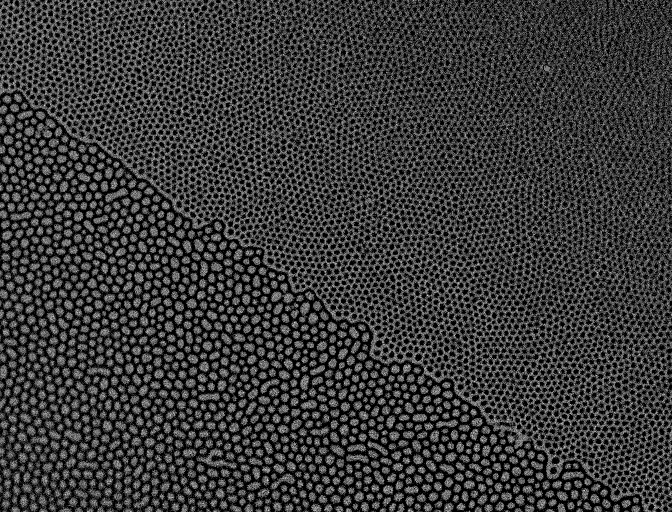
The images show exotic magnetic domains forming in Ta/CoFeB/MgO thin films after ion irradiation, ranging from labyrinth stripes to skyrmions and circles, even coexisting in different polarities and sizes.
UPK-01

Umut Parlak
Konstanz University
Postdoctoral Researcher
The image reveals magnetic domains formed within microscopic circular islands of a [Co/Pt] multilayer structure, ranging from 25 to 100 micrometers in diameter. Subjected to an alternating magnetic field applied perpendicular to the film plane, the material undergoes a delicate process of demagnetization. Rather than settling into disorder, the magnetization organizes itself into a sequence of concentric domains, creating rings that appear geometrically.
The materials were originally developed as a part of my PhD research project. However, these images were captured purely for aesthetic appreciation.
RKH-02

Ravinder Kaur
Indian Institute of Technology Hyderabad
PhD Candidate
Evolution of chiral spin textures in Ta(4)/Pt(4)/{Co(0.5)/Tb(0.65)/Co(0.5)}N/Pt(3) multilayers with increasing stack repetition (N=1–5). Room-temperature Kerr microscopy images reveal a morphological transition from magnetic bubble domains to isolated skyrmions as the number of Co/Tb/Co repetitions increases. The enhanced interfacial effects and dipolar interactions associated with larger N promote the stabilization of skyrmionic textures, demonstrating the crucial role of multilayer engineering in tailoring topological spin states.
CRH-01

Chennoju Raghu
Indian Insitute of Technology Hyderabad
PhD Candidate
Magneto-optical Kerr microscopy images showing the angular dependence of magnetization reversal in Co₂MnSi thin films. Due to the cubic crystalline anisotropy, domain wall motion varies with crystallographic direction. At 0° and 45°, magnetization switching occurs predominantly through the formation and propagation of band domains, indicating relatively easy reversal. In contrast, at 90° and 135°, the coexistence of band and ripple domains reflects increased anisotropy energy barriers and identifies these orientations as hard-axis directions. The domain evolution highlights the strong influence of magnetocrystalline anisotropy on the reversal mechanism.
SAN-01


Sikder Ashikuzzaman Ayon
Northwestern University
PhD Candidate
MOKE Imaging of Domain Wall Motion in Engineered Perpendicular Anisotropy Magnetic Multilayers for Memory Applications.
PFM-01

Piero Florio on behalf of PoliFab, PhyND Group, PoliMI
Politecnico di Milano
PhD Candidate
I am emotionally very attached to these images (they are among the first ever acquired with our Tr-MOKE instrument) and it was a great satisfaction to see spin waves propagating inside a magnonic circuit with my own eyes. Tr-MOKE (time-resolved magneto-optical Kerr effect) is a technique that captures snapshots of spin-wave dynamics by measuring the polarization rotation of a reflected pulsed laser beam; here the colormap represents the spin-wave phase.
a, Schematic of a programmable 2×2 phase-controlled router fabricated by direct laser writing (DLW). Two input spin waves are excited by a stripline antenna driven by an RF current and are then combined and routed according to their relative phase delay. The device consists of a phase shifter (red) cascaded with a directional coupler (orange).
b–d, Tr-MOKE phase maps showing phase-controlled routing. By tuning the phase shift and coupling parameter changing the external field, the initially in-phase spin waves are routed toward the top (b), bottom (d), or split equally between the two branches (c).
Scale bars: 10 µm.
APF-01

Alexej Perevertov
FZU - Institute of Physics, Prague
Staff Scientist
Magnetic domains in a Ni-Mn-Ga single crystal, imaged by wide-field Kerr microscopy with sensitivity aligned along the vertical direction (top image) and the horizontal direction (bottom image). The sample is in the martensitic state and is divided into twins with different orientations of the magnetic easy axis.
SBN-01

Sagarika Nayak on behalf of Bedanta etal., LNMM, NISER
B.J.B. Autonomous college, Bhubaneswar
Guest faculty
Role of interfacial exchange coupling on magnetic domains in hard/soft magnegtic bilayers: Magnetic domain imaging was performed on exchange-coupled soft/hard magnetic bilayer systems consisting of Co and (Co40Fe40B20) (CoFeB) by varying the thicknesses of the constituent layers. The first row of images corresponds to the Co(5 nm)/CoFeB(10 nm) bilayer, while the second row represents the Co(10 nm)/CoFeB(5 nm) bilayer. A significant modification in both the magnetic domain size and domain morphology was observed as a function of layer thickness. These changes arise from the interfacial exchange coupling between the soft magnetic Co layer and the relatively harder magnetic CoFeB layer. The results demonstrate that the magnetic domain configuration can be effectively tuned through thickness engineering, highlighting the crucial role of interfacial interactions in determining the magnetic behavior of exchange-coupled heterostructures. This work was carried out as part of my Ph.D. research under the supervision of Prof. Subhankar Bedanta at the National Institute of Science Education and Research (NISER), Bhubaneswar, India and was published in Journal of Physics D: Applied Physics 52 (2019) 305301.
SBN-04

Subhankar Bedanta on behalf of Prof. Subhankar Bedanta's lab
NISER-Bhubaneswar, India
Professor
Annealing-Induced Domain Evolution in in-plane anisotropic L10-ordered FeNi thin films -:Magneto-optical Kerr microscopy images showing the angular dependence of magnetization reversal in L10-ordered FeNi thin films with different anisotropy strengths. The images were recorded near the coercive field for field orientations of 0°, 30°, 60°, and 90°.As deposited sample exhibits large stripe domains that rotate with the applied field direction, indicating weak magnetic anisotropy and magnetization reversal dominated by domain-wall motion. After 100°C annealing displays narrower stripe domains together with the emergence of 90° domain walls, revealing the influence of cubic magnetic anisotropy. By further annealing of 100°C shows fine branched domains that remain largely unchanged with field orientation, reflecting an in-plane isotropic magnetic state with stronger effective anisotropy. The progressive reduction in domain width from Sample A to Sample C highlights the strong correlation between magnetic anisotropy and domain morphology, while the domain evolution confirms that magnetization reversal proceeds primarily through nucleation and domain-wall propagation. This work was carried out by my former postdoc Dr. V. Thiruvengadam at National Institute of Science Education and Research (NISER), Bhubaneswar. The study was published in Applied Physics Letters 115, 202402 (2019), providing important insights into the relationship between annealing, magnetic anisotropy, and domain evolution in L1₀-ordered FeNi thin films.
SBN-07

Mitali Swain on behalf of Prof. Subhankar Bedanta's Lab
National Institute of Science Education and Research Bhubaneswar, India
Postdoctoral Researcher
Microstructure-mediated magnetic behavior in NiFe-C60 bilayers : Investigated through magnetic domain imaging using longitudinal magneto-optical Kerr effect (LMOKE) microscopy at room temperature. Representative domain images are shown for S1: NiFe (20 nm), S2: NiFe (20 nm)/C60 (15 nm), and S3: C60 (15 nm)/NiFe (20 nm). The results demonstrate how stacking geometry influences interfacial hybridization in ferromagnet-organic semiconductor (FM-OSC) bilayers, with its impact clearly reflected in the magnetic domain evolution. For the single-layer NiFe film (S1), relatively larger magnetic domains and faster magnetization reversal are observed. In contrast, the bilayer samples (S2 and S3) exhibit significantly altered domain patterns and switching characteristics, indicating substantial modifications in the magnetic anisotropy energy landscape due to the presence of the C60 layer. Distinct domain behaviors observed in S2 and S3 highlight the crucial role of interface formation and deposition sequence in governing the magnetic properties of FM-OSC systems. These observations emphasize the importance of interface engineering for tailoring magnetic behavior in such systems. Furthermore, the study demonstrates the capability of Kerr microscopy as a powerful tool for visualizing and understanding the influence of microstructural factors such as interfacial hybridization, surface energy, strain, and defect distribution on magnetization reversal processes in thin-films. This work was carried out as part of my post doctoral work (DST/WOS-A/PM-43/2021) under the mentorship of Prof. Subhankar Bedanta at the National Institute of Science Education and Research (NISER), Bhubaneswar, India and was published in Journal of Physics D: Applied Physics 58 305303 (2025).
SBN-10

Srijani Mallik
Saha Institute of Nuclear Physics, Kolkata
Professor
Effect of spinterface on magnetic domain structure in ferromagnetic/organic semiconductor heterostructures: Longitudinal MOKE microscopy images illustrating the influence of the organic spinterface formed between a ferromagnet (Fe) and an organic semiconductor (C₆₀) on the magnetic domain structure. In our work published in Scientific Reports 8, 5515 (2018), we demonstrated that the non-magnetic fullerene C₆₀ acquires a measurable magnetic moment at the interface of an Fe (15 nm)/C₆₀ (40 nm) bilayer. This interfacial magnetization significantly modifies the magnetic domain structure and magnetization reversal process compared to a single Fe (15 nm) layer. For an epitaxial Fe thin film grown on MgO(001), magnetization reversal proceeds through the expected sequence of two 90° domain-wall motions, as shown on the left side of the dotted line. In contrast, the induced magnetic moment at the C₆₀/Fe spinterface alters the reversal mechanism, leading to multiple domain-wall propagation steps instead of the conventional two-step process, as illustrated on the right side of the dotted line. These observations provided direct evidence that organic spinterfaces can strongly influence the magnetic behavior of adjacent ferromagnetic layers. This study was one of the first to visualize the impact of an organic spinterface on the domain structure of ferromagnetic thin films. The work was carried out during my Ph.D. research under the supervision of Prof. Subhankar Bedanta at NISER, Bhubaneswar, India. Witnessing the emergence of unexpected domain patterns and unraveling the underlying physics was very exciting and intellectually rewarding for me.
LGS-02


Derek Bergner on behalf of Luo Lab
University of Southern California
PhD Candidate
Evidence of surface altermagnetism in altermagnetic candidate FeS. The ultra sensitive scanning Sagnac interferometer Polar MOKE microscope we employ is not only a mouthful but it can detect incredibly small Kerr rotations with a much higher sensitivity compared to conventional Kerr setups. Using this specialized instrument, we have been investigating the small uncompensated magnetic moments coming from the symmetry reduction due to a surface polishing of these crystals.
AMT-01

Carlos Garcia Garcia on behalf of Applied Magnetism and Spintronic Group
Universidad Tecnica Federico Santa Maria
Professor
Hysteresis loops measured by MOKE during the magnitude 7.1 earthquake that occurred in Valparaíso on April 24, 2017.
CME-01

Paul Keatley on behalf of Condensed Matter Group
University of Exeter
Facility Manager, The Exeter Time-Resolved Magnetism (EXTREMAG) Facility
A montage of counter-propagating spin waves in a 20 nm thick rectangle of Permalloy (Ni80Fe20). Spin waves were excited at the ends of the rectangle and propagate beautifully towards its centre. Using an in-plane 6 GHz microwave magnetic field excitation applied along the rectangle long edge, spin waves with different wavelength were isolated by tuning a static in-plane magnetic field applied along the short edge. The out-of-plane component of the spin wave precession was imaged using the polar magneto-optical Kerr effect in time-resolved scanning Kerr microscopy. High spatio-temporal resolution was achieved by focusing a pulsed laser with 140 fs pulse duration to a diffraction limited spot using x100 oil-immersion lens at wavelength of 520 nm. After almost 3 decades, time-resolved Kerr microscopy continues to be a powerful stroboscopic tool for exploring high-frequency and ultrafast magnetic processes in a wide range of magnetic materials and devices using the magneto-optical Kerr effects. Images were acquired in the EXTREMAG facility at the University of Exeter and reproduced courtesy of O. Barker, PhD Thesis, University of Liverpool (2026).
JGC-01


José Miguel García-Martín
CSIC
Research Scientist
Kerr domains observations for an epitaxial single phase Fe4N film which exhibits biaxial anisotropy in the plane due to its crystalline cubic structure. The field is applied horizontally (about 15° with respect to the closest easy axis), increasing in magnitude towards the left.
The images show the domain structure at the first magnetization transition driven by 90° domain walls (top row) followed by the second transition driven by another set of 90° domain walls (bottom row).
LLP-01

Louis Lerey
Paris-Saclay University
PhD Candidate
Temperature-dependent magnetic domain evolution in a Fe₃GaTe₂ flake imaged by polar MOKE microscopy.
Polar magneto-optical Kerr effect images of a large, flat and 8 nm (10 layers) thin exfoliated Fe₃GaTe₂ flake acquired at (a) 10°C, (b) 20°C, (c) 30°C, and (d) 35°C in the demagnetized state, revealing a stripe domain pattern with a well-defined up and down contrast arising from the strong perpendicular magnetic anisotropy (PMA) of this van der Waals ferromagnet. The domain period set by the competition between the magnetostatic energy and the domain wall energy decreases progressively with temperature. Thus it possible to retrieve some of the material's micromagnetic parameters from this temperature series.
Fe₃GaTe₂ is a room-temperature van der Waals ferromagnet with a Currie temperature of 350 K, that combines robust PMA, high spin polarization, and stable exfoliability down to few layers, making it a prime candidate for 2D spintronic and magnonic device integration.
Scale bar: each image is 40 µm wide.
SPB-01

Jean Carlos Rodriguez Estela on behalf of SPIN-TEAM Jean Lamour Institute
Instituto Balseiro, Universidad Nacional de Cuyo-CNEA, San Carlos de Bariloche, Río Negro, Argentina
PhD Candidate
The ghost of magnetic domains. Observation of a magnetic domain on a non-magnetic 100 nm copper layer. The magnetic contrast is due to the projection of an actual magnetic domain from a buried Co/Ni ferromagnetic multilayer. The image is obtained via a novel magneto-optical Kerr effect (MOKE) technique, namely the spin-accumulation based MOKE (SA-MOKE). The method uses optical excitations to remotely demagnetize the buried ferromagnetic layer to induce spin-pumping and a transient spin accumulation inside the Cu layer, resulting in magneto-optical contrast. The image size is 200 μm×200 μm.
IDB-02

Jean Carlos Rodriguez Estela on behalf of IDMAG - Laboratoire de Physique des Solides
Instituto Balseiro, Universidad Nacional de Cuyo-CNEA, San Carlos de Bariloche, Río Negro, Argentina
PhD Candidate
Bull’s-eye: nucleation and propagation. This figure shows the domain nucleation and magnetic wall propagation under high magnetic field pulses (pencil sketch). The images were carried out by PMOKE microscopy in [Tb(0.83nm)/Co(1.7nm)]x5 ferrimagnetic multilayers. The image size is 165 μm×165 μm.
YPB-02
Yi PENG
Beihang University
Postdoctoral Researcher
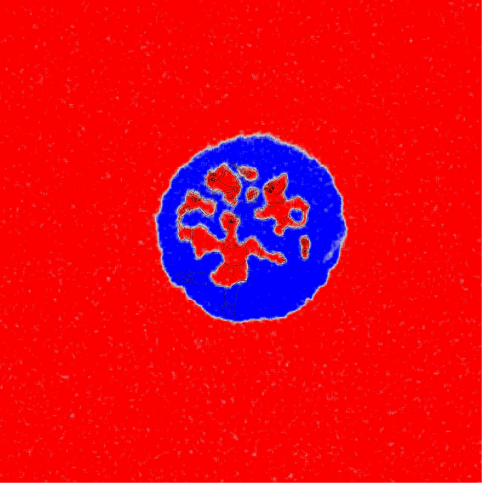
A magnetic dance after one femtosecond pulse
This Kerr microscopy image was recorded from a Co/Ho multilayer after excitation by a single 50-fs laser pulse. The red and blue regions represent opposite magnetization states, revealing the magnetic pattern written by the pulse.
Looking at the domain structure, I saw more than a switching pattern. Hidden in the Kerr contrast was a small scene: a girl dancing together with several animal-like companions. The line drawing highlights this personal visual interpretation. It is not intended as a scientific segmentation, but as a way to share the interesting visual poetry that can emerge from real magnetic domains . The original kerr image was published in Physical Review Applied 20, 014068 (2023).
TZB-01

Tianzhe ZHOU on behalf of Tianzhe ZHOU, Jingle CHEN, Weisheng ZHAO* and Nicolas VERNIER*
Beihang University and LuMIn, ENS Paris-Saclay.
MSc Candidate
Multilayer magnetic systems often exhibit complex magnetic configurations that cannot be fully resolved using a single imaging modality. To provide complementary magnetic information, we developed a home-built wide-field in-situ NV-MOKE microscope integrating polar Kerr microscopy and wide-field nitrogen-vacancy (NV) ensemble magnetometry. As shown in the top-centre panel, the system can be switched between NV and MOKE modes by simply engaging the flip mirror, polarizer, and analyzer. The lower panels illustrate the complementary measurement principles of the two modalities: MOKE imaging is primarily sensitive to the upper magnetic layer, whereas NV magnetometry measures the vector stray fields generated by the entire magnetic stack.
To demonstrate the capability of the NV-MOKE microscope, we investigated a multilayer sample consisting of Pt(1.5 nm)/Co(0.6 nm)/Ru(26.5 nm)/CoFeB(1.3 nm)/Ta(0.4 nm)/CoFeB(1.3 nm)/MgO(1.5 nm)/Ta(3 nm), containing two perpendicular magnetic anisotropy (PMA) layers. The upper-left MOKE image reveals the domain structure of the top Co layer, whereas the upper-right NV image contains stray-field contributions from both magnetic layers. Additional magnetic features observed in the NV image but absent from the MOKE image originate from the buried CoFeB layer.
The simultaneous acquisition of MOKE and NV images provides constraints for quantitative magnetic characterization. By combining domain structure information from MOKE with quantitative stray-field measurements from NV magnetometry, we can extract the saturation magnetization of the upper magnetic layer with a magnetic moment sensitivity down to 10⁻¹⁴ A·m² [Rev. Sci. Instrum. 96, 123711 (2025)]. Furthermore, once the magnetic state of the upper layer is determined, it is possible to reconstruct the magnetic configuration of the buried layer [In preparation].
NV-MOKE microscopy provides a combination of quantitative magnetic imaging and layer selectivity. While polar MOKE is primarily sensitive to out-of-plane magnetization, NV magnetometry probes the vector stray field and therefore contains information on both out-of-plane and in-plane magnetic components. It is thus possible to investigate multilayer magnetic systems with complex anisotropy configurations. More broadly, the capability to quantitatively resolve magnetic properties layer by layer without being restricted by magnetic anisotropy opens new possibilities for spatially resolved investigations of complex interlayer magnetic interactions in multilayer magnetic systems.
DYT-01


WARDA AL SAIDI on behalf of Dynaspin
University of Technology and Applied Sciences
Assistant Professor
The sample was a multilayer thin film with perpendicular magnetic anisotropy, designed to host complex domain configurations and possible topological magnetic textures. The goal was simple: measure the hysteresis behavior, image the domain evolution during field sweep, and understand how the magnetic state switched from one saturated direction to the other.
But the MOKE images revealed something far more interesting than a conventional reversal loop. During the positive branch of the hysteresis measurement, near the coercive field region, the film did not reverse uniformly. Instead of one clean switching event, the Kerr images showed a multilevel magnetic reversal. Different regions of the sample switched at different fields, forming several contrast levels in the Kerr images. These levels were not random noise; they represented distinct magnetic states appearing sequentially during the reversal process.
This observation changed the way we looked at the sample.
The hysteresis loop alone suggested a broader reversal process, but the MOKE images gave the hidden story behind it. They showed that the magnetization reversal was not a single-step transition, but a spatially resolved pathway involving domain-wall motion, localized delayed switching, and metastable magnetic configurations. Some regions reversed early, while others remained pinned and switched only later within a very narrow field window.
For me, this was the moment when the Kerr image became more than a visualization tool. It became a way to listen to the magnetic material after the external field had been removed. The most important part of the experiment was not only the hysteresis loop, but the backstory hidden between its branches: the delayed switching, the pinned domains, the metastable regions, and the slow magnetic relaxation at zero field. Without MOKE imaging, this multilevel reversal would have appeared only as a broadened or sheared hysteresis curve. With MOKE, we could see the magnetic process unfold directly in space and time.
These images opened a new direction in our research. They suggested that the material may support complex multistate magnetic behavior, where different domain configurations can be stabilized, delayed, or transformed depending on magnetic history. This is especially relevant for future spintronic and neuromorphic devices, where information could be encoded not only in binary magnetic states, but also in intermediate, history-dependent magnetic configurations.
RKH-03

Ravinder Kaur
Indian Institute of Technology Hyderabad
PhD Candidate
Room-temperature Kerr microscopy image of GdCo near its magnetic compensation point under an out-of-plane magnetic field. A mixed magnetic phase comprising labyrinth domains and skyrmions is observed, where the labyrinthine stripes progressively fragment into isolated skyrmions. This coexistence reflects the field-driven transition between extended chiral domains and topologically stabilized spin textures.
LZW-01

Le Zhao
TU Wien
Postdoctoral Researcher
These MOKE images capture geometry-induced magnetic domain patterns in Ta/CoFeB/MgO/Ta multilayers grown on Si/SiO₂ substrates with shallow etched indentations. The substrate was first patterned with periodic squares, hexagons, triangles, and other recessed shapes, only about 6 to 10 nanometers deep. After deposition, this subtle topography was transferred into the magnetic multilayer, creating local variations in the nucleation energy landscape. The result is a set of rich artificial magnetic domain patterns, where the etched geometry quietly guides how domains first appear and evolve. This was the last project of my PhD at Tsinghua University and was eventually published in Science Bulletin (https://doi.org/10.1016/j.scib.2024.05.035). More importantly, it changed the way I thought about magnetic thin films. I began to see geometry not merely as a fabrication detail, but as an active parameter that can control magnetic states. That realization became one of the reasons I later moved toward the study of three-dimensional and non-planar magnetic structures, eventually joining TU Wien as a postdoctoral researcher to continue exploring this direction.
SAN-02

Sikder Ashikuzzaman Ayon
Northwestern University
PhD Candidate
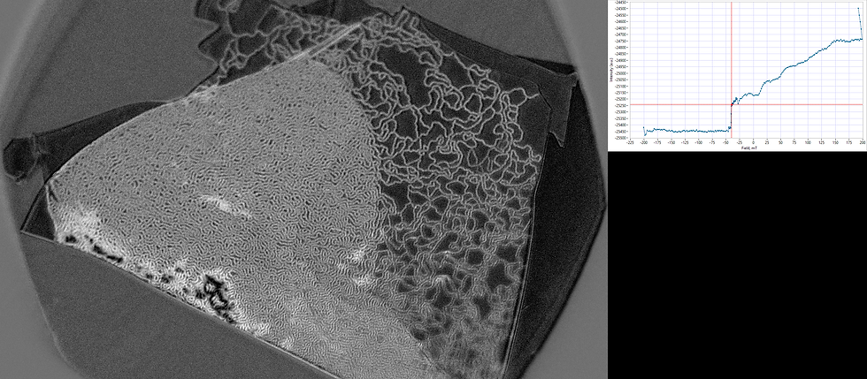
Resolving the magnetic domains in Fe3GaTe2 at room temperature.
ISD-01

Ivan Soldatov
IFW-Dresden
Staff Scientist
Magnetic domains in a metallic multilayer thin film with DMI, imaged in an AC-demagnetized state. The sample was placed between two iron pole pieces such that the sample plane aligned with the top surface of the poles. This configuration forces the perpendicular component of the magnetic field to change its sign directly in the center of the sample. The image was acquired using a wide-field Kerr microscope optimized for polar sensitivity.
SMS-01

Sougata Mallick
SRM Institute of Science and Technology, Kattankulathur, India
Professor
Polar MOKE microscopy images illustrating domain engineering via magnetic antidot lattices in a PtICo multilayer system. Panels (a–c) correspond to the domain images for the Pt|Co continuous film, while panels (d–f) show the same magnetic system microfabricated with a periodic array of triangular antidots (hole size ~10 μm). Images were recorded at the saturation (a and d), nucleation (b and e), and coercive fields (c and f). The direction of the applied magnetic field and the scale bar are identical for all images and are shown in panel (a). The insets in panels (e) and (f) present magnified views of the regions highlighted by the yellow boxes.
A direct comparison between the continuous and antidot-patterned films reveals pronounced domain engineering induced by the artificial pinning landscape and geometrical constrictions. The average domain sizes for the continuous thin film and antidot lattices are ~100 μm and ~10 μm, respectivelty. This work was part of my PhD thesis work under the supervision of Prof. Subhankar Bedanta at NISER, Bhubaneswar, India. I was very happy to observe these domain images which evidence the importance of antidot lattices for future spintronic applications. This work was published in Scientific Reports 8, 11648 (2018).
SBN-02

Abhisek Mishra on behalf of Prof. Subhankar Bedanta's lab
NISER Bhubaneswar, India
Postdoctoral Researcher
Hybridization induced anisotropy in Nanoscale-Thick MoS₂/CoFeB Heterostructures: Magneto-optical Kerr effect (MOKE) microscopy images recorded in the longitudinal geometry at room temperature with the magnetic field applied at angles of 0°, 45°, and 90°. Panels (a–c) show the magnetic domain structures of the bare CoFeB (6 nm) thin film, while panels (d–f) correspond to the CoFeB (6 nm)/MoS₂ (28 nm) heterostructure. The scale bar shown in panel (a) is applicable to panels (b) and (c), whereas the scale bar in panel (d) is valid for panels (e) and (f). The magnetic field orientation indicated in panel (a) applies to all images. These results were published in ACS Applied Nano Materials 5, 10645 (2022).
The domain evolution provides direct microscopic evidence of the magnetic anisotropy modification induced by the MoS₂ underlayer. The bare CoFeB film exhibits nearly isotropic domain behavior, whereas the CoFeB/MoS₂ heterostructure displays pronounced changes in domain structures, indicative of enhanced magnetic anisotropy. This enhancement is attributed to interfacial hybridization between the electronic states of MoS₂ and the Co/Fe atoms at the interface highlighting the crucial role of interface engineering in tailoring magnetic properties. This work formed an important part of my Ph.D. research conducted under the supervision of Prof. Subhankar Bedanta at NISER Bhubaneswar, India. Observing such distinct domain transformations was particularly rewarding, as it provided direct visual evidence of how interfacial interactions can be utilized to tune and manipulate magnetic anisotropy, offering promising pathways for the design of energy-efficient spintronic technologies.
SBN-05

Brindaban Ojha on behalf of Prof. Subhankar Bedanta's lab
Uppsala University
Postdoctoral Researcher
Unusual domain wall motion in the vicinity of the depinning field in a Pt/CoFeB/MgO film: Bubble domain images of an out-of-plane magnetized Pt/CoFeB/MgO system measured using polar magneto-optical Kerr effect (MOKE) microscopy. (a) Differential domain images obtained by subtracting consecutive images, where the inner and outer boundaries represent the nucleated and propagated domains, respectively. (b) Domain wall (DW) velocity as a function of the applied magnetic field, calculated from the DW displacement over the time interval between successive images. The DW dynamics exhibit an "excess velocity" regime compared to the conventional creep law behaviour. This enhancement is attributed to additional relaxation events occurring near the depinning field, as discussed in detail in Ref. [Appl. Phys. A 129, 688 (2023)]. This work was carried out as part of my Ph.D. research under the supervision of Prof. Subhankar Bedanta at the NISER, Bhubaneswar, India. The measurements are performed at LPS, Université Paris-Saclay, France.
SBN-08

Purbasha Sharangi on behalf of Prof. Subhankar Bedanta's lab
Istituto Nazionale di Ricerca Metrologica (INRIM), Italy
Postdoctoral Researcher
Role of spinterface on magnetization reversal in a perpendicularly magnetic anisotropic system: Polar MOKE microscopy images illustrating domain engineering induced by spinterface formation in perpendicularly magnetized Pt/Co/C_60/Pt heterostructures. Panels (a–e) show domain evolution in the Pt/Co/Pt reference sample, while panels (f–j), correspond to Pt/Co/C_60/Pt heterostructures with C_60 thicknesses of 3.2 nm, respectively. Images were recorded at positive saturation (a and f), near nucleation (b and g), between nucleation and coercive field (c and h), at coercive field (d, and i) and near negative saturation (e, and j). The direction of the applied magnetic field and the scale bar are identical for all images and are indicated in panel (a). For better visualization, domains are marked as red circles and the diameter of the circles is the same for (b and g) and (c and h).
A direct comparison of the perpendicularly magnetized Pt/Co/Pt and Pt/Co/C_60/Pt heterostructures with varying C_60 thicknesses reveals significant domain engineering arising from spinterface formation. The introduction of a C_60 interlayer into the Pt/Co/Pt thin film leads to a reduction in domain size. This work was part of my PhD thesis work under the supervision of Prof. Subhankar Bedanta at NISER-Bhubaneswar, India. I was delighted to observe these domain images, which clearly demonstrate the ability to engineer domain size through tuning of the spinterface thickness. These results have been published in J. Mater. Chem. C 10, 17236 (2022).
SBN-11

Shaktiranjan Mohanty on behalf of Prof. Subhankar Bedanta's Lab
NISER, Bhubaneswar, India
Postdoctoral Researcher
Magnetization reversal and domain evolution in synthetic antiferromagnets with perpendicular magnetic anisotropy:
Polar MOKE microscopy images reveal how antiferromagnetically coupled [Co/Pt]/Ir/[Co/Pt] multilayers reverse their magnetization through distinct domain nucleation and domain-wall propagation processes. (a) and (b) show the corresponding polar MOKE hysteresis loops, while figures (a1–a4), (b1–b4), present domain images recorded at the magnetic field values marked on the respective loops. Although all samples exhibit antiferromagnetic interlayer coupling, their domain structures differ significantly, ranging from nearly homogeneous reversal to the formation of large reversed domains and multi-step switching. These observations provide direct visual evidence of how layer thickness and interlayer exchange coupling govern the reversal pathways in synthetic antiferromagnets with perpendicular magnetic anisotropy. The results were published in JOM 74, 2319-2327 (2022). This work was carried out during my Ph.D. research under the supervision of Prof. Subhankar Bedanta at NISER Bhubaneswar, India. Observing the correlation between Kerr domain images and complex hysteresis-loop shapes was particularly exciting, as it helped uncover the underlying magnetization reversal mechanisms in these SAF systems.
AMC-01

Andrea Migliorini
Ca' Foscari University of Venice
Assistant Professor
A Kerr-microscope snapshot taken while the antiferromagnetic IrMn layer of a FeCo/IrMn bilayer crystallizes, the moment a structural phase transition spontaneously writes exchange bias into the film. The large image is a single Kerr field of view, shown as raw contrast on one side and colour-coded by region on the other. The bull's-eye rings are frozen history: as the IrMn layer crystallised, the in-plane FeCo remanence was switched one way, then the other, then back again, and each state was pinned in turn, from the inside out. The VSM loops (i to iv) follow the growing biased fraction, with the marker flagging the state of each sketch. Left to itself, the outer biased ring would keep spreading until it claimed the whole sample.
What I love about it: the exchange bias set itself, and Kerr microscopy let us watch it switch on, ring by ring, as the crystal formed.
Originally published in Nature Materials 17, 28–35 (2018) (DOI: 10.1038/nmat5030) during my PhD in Prof. José Luis Prieto's group at the Universidad Politécnica de Madrid, in collaboration with Julio Camarero's group at IMDEA Nanociencia and Kevin O'Grady's group at the University of York.
Cast Your Vote
Once the mosaic is assembled, the magnetics community will vote for their favorite entries. Voting opens in early July 2026 and closes in September 2026 (updated). Each person/group is only eligible for one award, regardless of the total number of submissions by the individual/group.
Winning entries will receive special recognition and prizes (details to be announced).
Entries submitted late will not be considered for voting.
How to Vote
Each entry in the Kerr 150 mosaic is labeled with an index number (e.g., ABC-01, DEF-03) at the top. Use this index number to identify your favorites, and input it into the form below.
(Tip: copy and paste the index into the relevant boxes in the form!)
What to vote for: Choose the entries you find most inspiring, interesting, or creative — submissions that best celebrate the spirit and beauty of the Kerr effect. This isn't strictly a technical merit contest.
You may vote for up to 3 entries. List them in order of preference.
Voting closes on 14 September 2026 (Monday) 2359 Eastern Time.
Voter Eligibility & Fair Play
To keep voting fair and reflective of genuine community sentiment, please note:
-
Votes must come from the voter's active email address, registered to a recognized institutional or organizational affiliation (no personal/anonymous burner emails, e.g. @gmail.com, @yahoo.com, etc.).
-
One vote per person. Submitting multiple times will result in disqualification of all your votes.
-
Voters should not vote for their own submission(s) or those from the research group/lab(s) they are associated with presently and/or previously, to avoid conflicts of interest.
-
Organizers reserve the right to disregard votes where coordinated bloc voting or other suspicious activity is suspected, at their discretion.
-
These safeguards exist to protect the spirit of the event and ensure fairness to all participants and contributors.
-
Voting is one input, not the sole determinant. A panel of judges from the magnetics community will also evaluate entries, and final prize decisions will combine community votes with judges' assessments.
-
By submitting the voting form, you agree to provide accurate affiliation information; organizers may verify affiliations against submitter records.
Voting Form

[Closed] Submit Your Contributions
For multiple images with different captions, please submit them separately. Each form submission will only be tagged to one caption.
Feel free to submit as a research group!
As written in the submission form:
-
The coordinators of this event reserve the right to remove any material they deem offensive, inappropriate, or unsuitable for the context of the event.
-
By submitting the information provided by the participants onto the forms, participants acknowledge that:
-
they hold the right to share the images(s) and information provided in the form. SiM and IEEE Magnetics Society takes no responsibility for the accuracy, completeness, or publication status of submitted content. Submitting unpublished data remains the sole responsibility of the contributor.
-
they acknowledge and consent to having my submission featured publicly on the IEEE Magnetics Society platforms.
-
Acknowledgements
This event is a joint initiative by SiM, IEEE Magnetics Society Young Professionals, and the INTERMAG 2026 Kerr Symposium.
